The Synapse
This is the longest and most tedious module. Don't overthink the vocab.
Covers: Basic vocabulary of brain signaling.
The Synapse
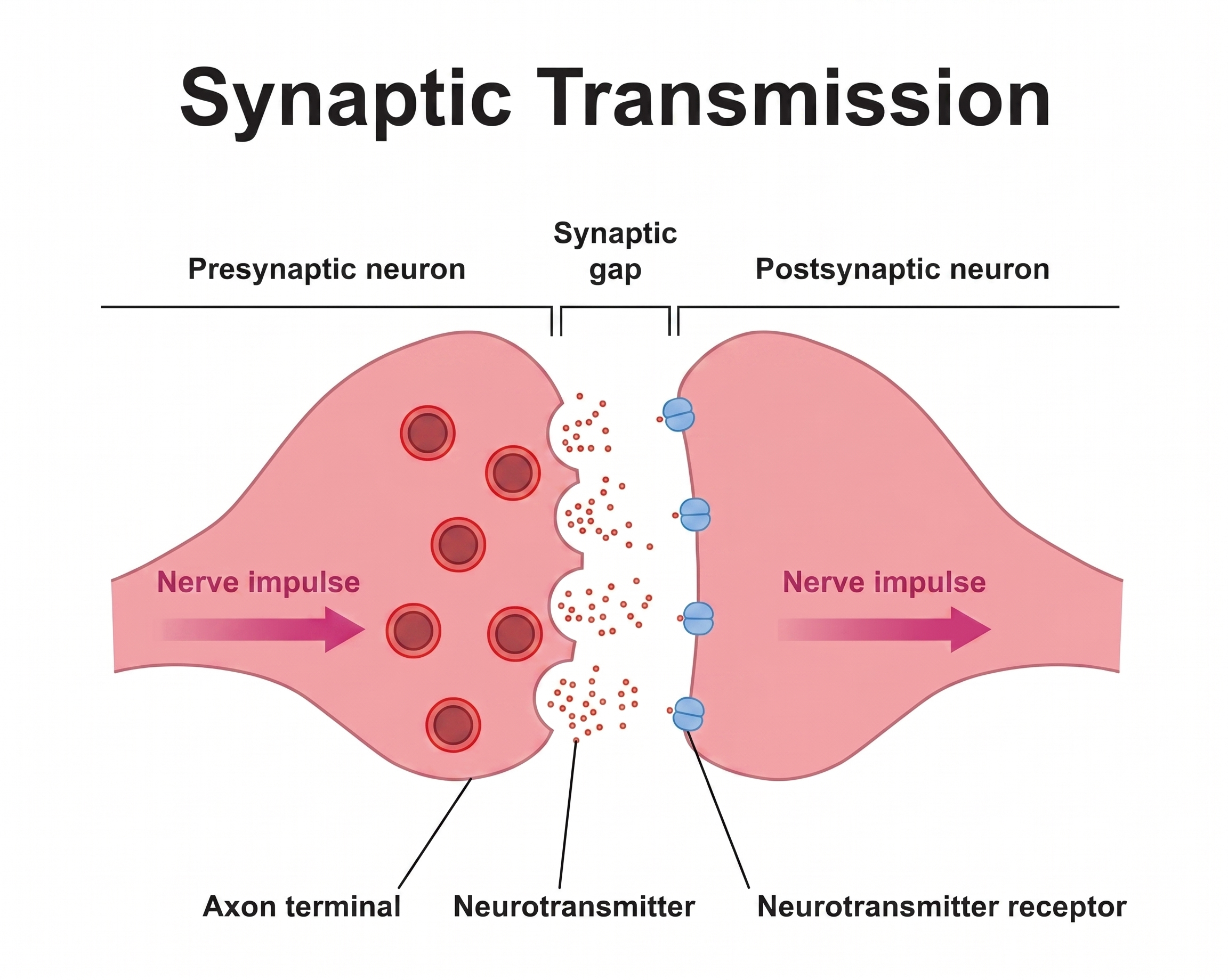
The synapse is the gap between two nerve cells. Three parts matter:
- Presynaptic neuron - the sender. Stores neurotransmitters in small packets called vesicles. When an electrical signal arrives, those packets fuse with the membrane and dump their contents into the gap.
- Synaptic cleft - the empty space the chemicals must cross.
- Postsynaptic neuron - the receiver. Has receptors. When the right chemical binds to a receptor, something happens. If nothing fits, nothing happens.
Video: Synaptic Transmission ↗ YouTube
How drugs work: they do something to the message. They mimic neurotransmitters, block receptors, prevent cleanup, or force a dump.
The Ions: How Neurons Actually Fire
Neurons fire by moving charged ions across their cell membrane. There are 4 main ions for this course.
-
Sodium (Na⁺) - the "go" ion.
- Normally concentrated outside the neuron.
- When sodium channels open, Na⁺ rushes in → inside becomes more positive → depolarization (more likely to fire).
- Without sodium influx, the neuron cannot send a signal.
-
Potassium (K⁺) - the "reset" ion.
- Normally concentrated inside the neuron.
- When potassium channels open, K⁺ rushes out → inside becomes more negative → hyperpolarization (less likely to fire).
-
Chloride (Cl⁻) - the "quiet" ion.
- Normally concentrated outside the neuron.
- When chloride channels open, Cl⁻ rushes in → inside becomes more negative → hyperpolarization.
-
Calcium (Ca²⁺) - the "release" ion.
- Normally concentrated outside the neuron.
- When voltage-gated calcium channels open at the presynaptic terminal, Ca²⁺ rushes in → triggers vesicles to fuse with the membrane → neurotransmitter dumps into the synapse.
The Action Potential
The action potential is the electrical signal that travels down a neuron. The sequence:
- At rest, the inside of the neuron sits at around −70 mV (more negative than outside).
- A trigger pushes the membrane voltage upward.
- If the trigger crosses a threshold (~−55 mV), voltage-gated sodium channels open → Na⁺ rushes in → voltage shoots up to ~+30 mV.
- Potassium channels open → K⁺ rushes out → voltage drops back down.
- The wave moves down the axon to the terminal.
- At the terminal, depolarization opens voltage-gated calcium channels → Ca²⁺ enters → vesicles release neurotransmitter into the synapse.
- Threshold membrane potential (~−55 mV) - The voltage a neuron must reach to fire a full action potential.
- Below threshold, any depolarization is sub-threshold: the neuron gets pushed closer to firing but doesn't commit.
- Partial depolarization is what some drugs cause: they nudge the membrane toward threshold without crossing it.
- At threshold, the response is all-or-nothing.
Video: Action Potential ↗ YouTube
One-line summary: Sodium fires it, potassium and chloride quiet it, calcium causes the release of the chemical signal.
Receptor Types
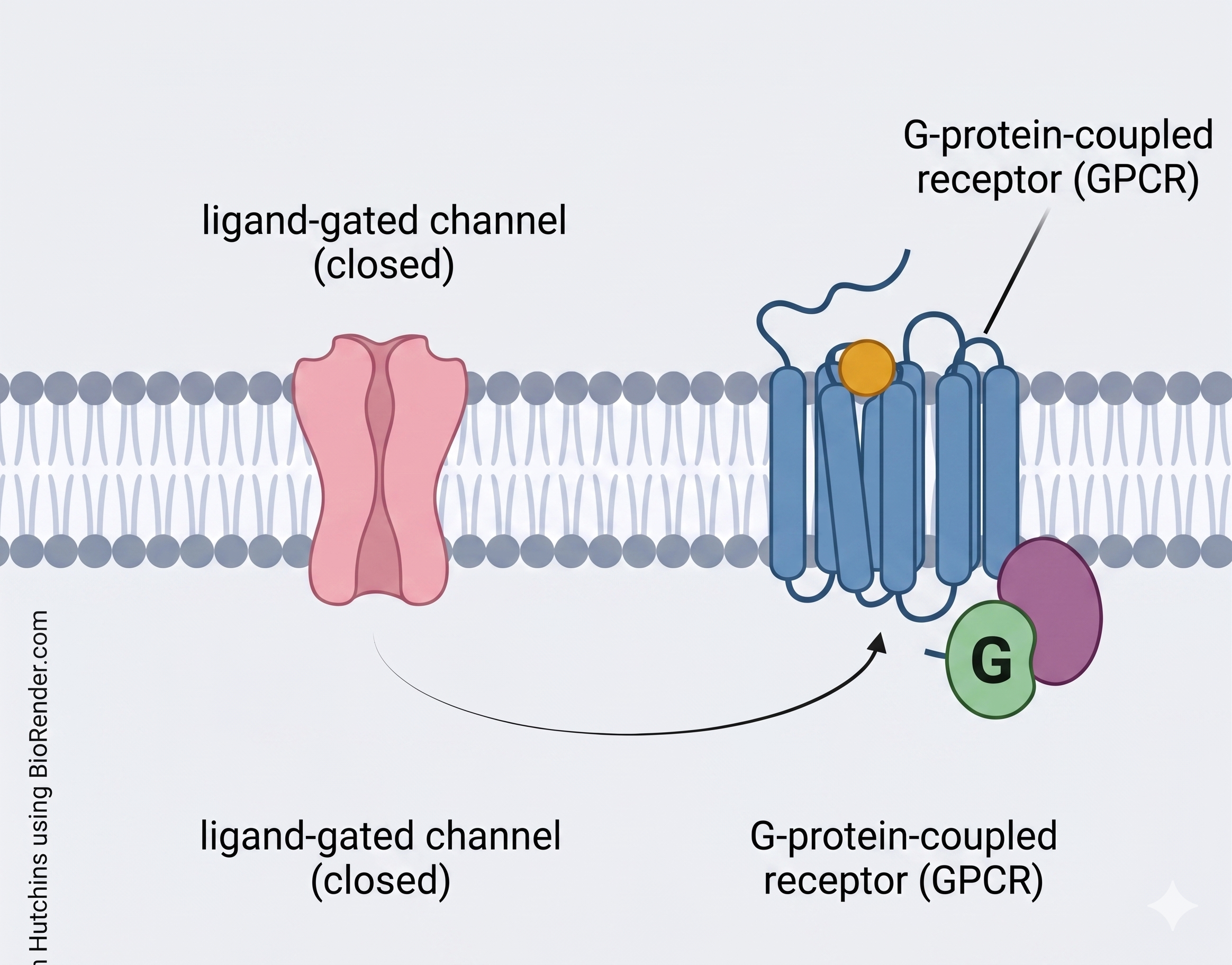
- Metabotropic or G-protein coupled receptors (GPCRs) - neurotransmitter binds → triggers an internal signaling cascade via the G-protein.
- Slower response, produces broader and longer-lasting cellular changes.
- Ionotropic receptors (Ligand gated ion channels) - something binds → directly opens an ion channel.
- Quicker response that immediately changes the voltage of the neuron.
Agonists and Antagonists
- Agonist - binds to a receptor and activates it, producing a full biological response equal to the body's natural signaling molecule.
- Partial Agonist - binds to a receptor and activates it, but only produces a partial (submaximal) response even when all receptors are occupied. When a full agonist is also present, a partial agonist can act as a net blocker by competing for the same binding sites.
-
Antagonist - binds to a receptor but does not activate it at all. Prevents agonists from binding, effectively blocking or dampening the biological response.
- Naloxone is a competitive antagonist at the mu opioid receptor (MOR) - it blocks opioid binding without activating the receptor, which is how it reverses overdose.
-
Inverse Agonist - binds to a receptor and decreases its baseline (constitutive) activity. Produces a response that is the exact opposite of an agonist: rather than just blocking action, it actively suppresses it.
- Beta-blockers (e.g., propranolol, carvedilol) act as inverse agonists at beta-adrenergic receptors, actively suppressing constitutive receptor activity rather than simply blocking it. This is thought to contribute to their beneficial effects in heart failure (Yoshikawa, 2005).
Video: Receptors & Ligands ↗ YouTube
Second Messengers: cAMP
When a neurotransmitter binds a receptor, sometimes it opens an ion channel directly (fast, within milliseconds). Other times it activates an internal signaling cascade that produces longer-lasting changes (seconds to hours).
Cyclic adenosine monophosphate (cAMP) is the most important second messenger to know for drugs. It is made inside the cell by an enzyme called adenylyl cyclase. cAMP translates external signals (like neurotransmitters binding to receptors) into specific cellular responses.
Many drug receptors are coupled to cAMP. When the drug binds, cAMP levels go up or down and the neuron's behavior shifts over hours, days, or weeks. This is the cellular mechanism behind tolerance and withdrawal.
Decreased cAMP → Cell becomes less active.
Increased cAMP → Cell becomes more active.
Video: Second Messenger cAMP - click to expand ↗ YouTube
Video: cAMP Signaling in depth - click to expand ↗ YouTube
Click on underlined words throughout the course to see their definition. Over time you'll know them. They come up constantly.
Sources
- Biology Blues. (n.d.). Second messenger cAMP [Video]. YouTube. https://www.youtube.com/watch?v=GhOFkWWVqp4
- Neuroscientifically Challenged. (n.d.). 2-Minute neuroscience: Action potential [Video]. YouTube. https://www.youtube.com/watch?v=W2hHt_PXe5o
- Neuroscientifically Challenged. (n.d.). 2-Minute neuroscience: Receptors & ligands [Video]. YouTube. https://www.youtube.com/watch?v=NXOXZ-kaSVI
- Neuroscientifically Challenged. (n.d.). 2-Minute neuroscience: Synaptic transmission [Video]. YouTube. https://www.youtube.com/watch?v=WhowH0kb7n0
- Rethink Biology. (n.d.). GPCR cAMP signaling || Second messenger cAMP || 4K animation [Video]. YouTube. https://www.youtube.com/watch?v=XIdBJm8DaFo
- Yoshikawa, T. (2005). Inverse agonism of beta-blockers. Journal of Cardiac Failure, 11(9), S244. https://doi.org/10.1016/j.cardfail.2005.08.045